Tests are routinely used to detect early stages of some types of cancer (such as breast, cervical, colorectal, and skin cancer) before they cause symptoms. At this time, no special tests are routinely recommended to detect bone cancers early. The best strategy for early diagnosis is prompt attention to the signs and symptoms of this disease.
Signs and Symptoms of Bone Cancer
Pain
Pain in the affected bone is the most common complaint of patients with bone cancer. At first, the pain is not constant. It may be worse at night or when the bone is used (for example, leg pain when walking). As the cancer grows, the pain will be there all the time. The pain increases with activity and the person might limp if a leg is involved.
Swelling
Swelling in the area of the pain may not occur until weeks later. It might be possible to feel a lump or mass depending on the location of the tumor.
Cancers in the bones of the neck can cause a lump in the back of the throat that can lead to trouble swallowing or make it hard to breathe.
Fractures
Bone cancer can weaken the bone it develops in, but most of the time the bones do not fracture (break). People with a fracture next to or through a bone cancer usually describe sudden severe pain in a limb that had been sore for a few months.
Other Symptoms
Cancer in the bones of the spine can press on nerves, leading to numbness and tingling or even weakness.
Cancer can cause weight loss and fatigue. If the cancer spreads to internal organs it may cause other symptoms, too. For example, if the cancer spreads to the lung, you may have trouble breathing.
Any of these symptoms are more often due to conditions other than cancer, such as injuries or arthritis. Still, if these problems go on for a long time without a known reason, you should see your doctor.
How is Bone Cancer Diagnosed?
A patient’s symptoms, physical exam, and results of imaging tests, and blood tests may suggest that bone cancer is present. But in most cases, doctors must confirm this suspicion by examining a tissue or cell sample under a microscope (a procedure known as a biopsy).
Other diseases, such as bone infections, can cause symptoms and imaging results that could be confused with bone cancer. Accurate diagnosis of a bone tumor often depends on combining information about its location (what bone is affected and even which part of the bone is involved), appearance on x-rays, and appearance under a microscope.
Since a single bone metastasis can have the same signs and symptoms as a primary bone tumor, many doctors require a biopsy to diagnose a patient’s first bone metastasis. After that, additional bone metastases can usually be diagnosed based on x-rays and other imaging tests.
Imaging Tests to Detect Bone Cancer
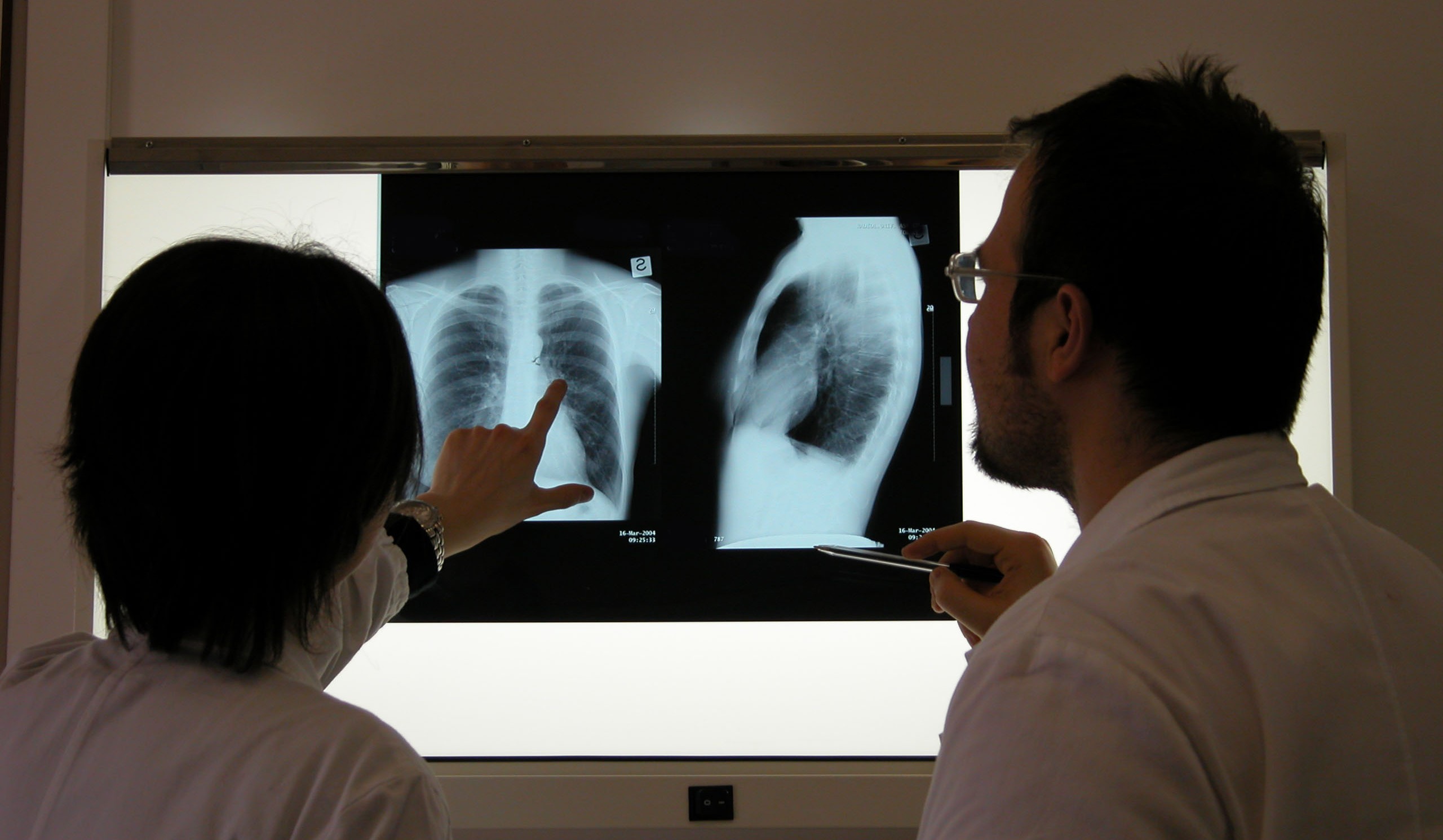
X-Rays
Most bone cancers show up on x-rays of the bone. The bone at the site of the cancer may appear “ragged” instead of solid. The cancer can also appear as a hole in the bone. Sometimes doctors can see a tumor around the defect in the bone that might extend into nearby tissues (such as muscle or fat). The radiologist (doctor who specializes in reading x-rays) can often tell if a tumor is malignant by the way it appears on the x-ray, but only a biopsy can absolutely determine that.
A chest x-ray is often done to see if bone cancer has spread to the lungs.
Computed Tomography (CT) Scans
The CT scan is an x-ray procedure that produces detailed, cross-sectional images of your body. Instead of taking one picture, like a conventional x-ray, a CT scanner takes many pictures as it rotates around you. A computer then combines these pictures into an image of a slice of your body. The machine creates multiple images of the part of your body that is being studied.
A CT scanner has been described as a large donut, with a narrow table in the middle opening. You will need to lie still on the table while the scan is being done. CT scans take longer than regular x-rays, and you might feel a bit confined by the ring while the pictures are being taken.
CT scans are helpful in staging cancer. They help tell if your bone cancer has spread into your lungs, liver, or other organs. These scans also show the lymph nodes and distant organs where metastatic cancer might be present.
Before the test you may be asked to drink 1 or 2 pints of a contrast agent. This helps outline the stomach and intestine to make it easier to see tumors. You might also receive an IV (intravenous) line through which a different kind of contrast dye is injected. This helps better outline structures in your body.
The injection can cause some flushing (redness and warm feeling that may last hours to days). A few people are allergic to the dye and get hives. Rarely, more serious reactions like trouble breathing and low blood pressure can occur. You can be given medicine to prevent and treat allergic reactions. Be sure to tell the doctor if you have ever had a reaction to any contrast material used for x-rays or if you have an allergy to shellfish.
CT scans can also be used to precisely guide a biopsy needle into a suspected metastasis. For this procedure, called a CT-guided needle biopsy, the patient remains on the CT scanning table while a radiologist advances a biopsy needle toward the location of the mass. CT scans are repeated until the doctors are confident that the needle is within the mass. (See the section, “Needle biopsy.”)
Magnetic Resonance Imaging (MRI) Scans
MRI scans use radio waves and strong magnets instead of x-rays. The energy from the radio waves is absorbed and then released in a pattern formed by the type of tissue and by certain diseases. A computer translates the pattern of radio waves given off by the tissues into a very detailed image of parts of the body. Sometimes a contrast material called gadolinium is injected into your veins to better see the tumor.
MRI scans are often the best test for outlining a bone tumor. They are also particularly helpful for looking at the brain and spinal cord. MRI scans are a little more uncomfortable than CT scans. First, they take longer — often up to an hour. Also, you have to be placed inside a tube, which is confining and can upset people with claustrophobia (fear of enclosed spaces). The machine also makes a thumping noise that you may find disturbing. Some places provide headphones with music to block this out.
Radionuclide Bone Scans
This procedure helps show if a cancer has spread to other bones. It can find metastases earlier than regular x-rays. Bone scans also can show how much damage the primary cancer has caused in the bone.
For this test, the patient receives an injection of radioactive material called technetium diphosphonate (tek-NEE-shee-um die-fos-fuh-nate). The amount of radioactivity used is very low and causes no long-term effects. This substance is attracted to diseased bone cells throughout the entire skeleton. Areas of diseased bone will be seen on the bone scan image as dense, gray to black areas, called “hot spots.” These areas suggest metastatic cancer is present, but arthritis, infection, or other bone diseases can also cause a similar pattern. To distinguish among these conditions, the cancer care team may use other imaging tests or take bone biopsies.
Positron Emission Tomography (PET or PET) Scans
PET scans use glucose (a form of sugar) that contains a radioactive atom. A special camera can detect the radioactivity. Cancer cells absorb a lot of the radioactive sugar because of their high rate of metabolism. PET scans are useful in looking for cancer throughout your entire body. It can sometimes help tell if a tumor is cancerous or benign. It is being combined with CT scans to better pinpoint some kinds of cancer.
Biopsy
A biopsy is a sample of tissue taken from a tumor so that it can be looked at under a microscope. This is the only way to know that the tumor is cancer and not some other bone disease. If cancer is present, the biopsy can tell the doctor if it is a primary bone cancer or cancer that started somewhere else and spread to the bone (metastasis). Several types of tissue and cell samples are used to diagnose bone cancer. It is very important a surgeon with experience in diagnosing and treating bone tumors do the biopsy procedure.
The surgeon will choose a biopsy method based on whether the tumor looks benign or malignant and exactly what type of tumor is most likely (based on the bone x-rays, the patient’s age, and the location of the tumor). Some kinds of bone tumors can be recognized from needle biopsy samples, but larger samples (from a surgical biopsy) are often needed to diagnose other types. Whether the surgeon plans to remove the entire tumor at the time of the biopsy will also influence the choice of biopsy type. The wrong kind of biopsy can sometimes make it hard later for the surgeon to remove all of the cancer without having to also remove all or part of the arm or leg containing the tumor. It also may cause the cancer to spread.
Needle Biopsy
There are 2 types of needle biopsies: fine needle biopsies and core needle biopsies. For both types, a local anesthetic is first used to numb the area for the biopsy. For fine needle aspiration (FNA), the doctor uses a very thin needle attached to a syringe to withdraw a small amount of fluid and some cells from the tumor mass. Sometimes, the doctor can aim the needle by feeling the suspicious tumor or area that is near the surface of the body. If the tumor cannot be felt because it is too deep, the doctor can guide the needle while viewing a CT scan. This is called a CT guided needle biopsy and it is often done by an x-ray specialist known as an interventional radiologist. In a core needle biopsy, the doctor uses a larger needle to remove a small cylinder of tissue (about 1/16 inch in diameter and 1/2 inch long). Many experts feel that a core needle biopsy is better than FNA to diagnose a primary bone cancer.
Surgical Bone Biopsy
In this procedure, a surgeon needs to cut through the skin to reach the tumor in order to remove a small piece of tissue. This is also called an incisional biopsy. If the entire tumor is removed (not just a small piece), it is called an excisional biopsy. These biopsies are often done with the patient under general anesthesia (asleep). They can also be done using a nerve block, which numbs a large area. If this type of biopsy is needed, it is important that the surgeon who will later remove the cancer also be the one to do the biopsy.
Source: http://www.cancer.org/