Early detection improves the chances that male cancer can be treated successfully.
Differences Affecting Early Detection of Male and Female Breast Cancers
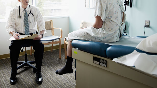
There are many similarities between breast cancer in men and women, but there are some important differences that affect finding it early.
Breast Size
The most obvious difference between the male and female breast is size. Because men have very little breast tissue, it is easier for men and their health care professionals to feel small masses (tumors). On the other hand, because men have so little breast tissue, cancers do not need to grow very far to reach the nipple, the skin covering the breast, or the muscles underneath the breast. So even though breast cancers in men tend to be slightly smaller than in women when they are first found, they more often have already spread to nearby tissues or lymph nodes. The extent of spread is one of the most important factors in the prognosis (outlook) of a breast cancer.
Lack of Awareness
Another difference is that breast cancer is common among women and rare among men. Women tend to be aware of this disease and its possible warning signs, but many men do not think that they can get it at all. Some men ignore breast lumps or think they are caused by an infection or some other reason, and they do not get medical treatment until the mass has had a chance to grow. Some men are embarrassed when they find a breast lump and worry that someone might question their masculinity. This could also delay diagnosis and reduce a man’s chances for successful treatment.
Because breast cancer is so uncommon in men, there is unlikely to be any benefit in screening men in the general population for breast cancer with mammograms or other tests.
For Men Who Are or May Be at High Risk
Careful breast exams might be useful for screening men with a strong family history of breast cancer and/or with BRCA mutations found by genetic testing. Screening men for breast cancer has not been studied to know if it is helpful, and mammography (x-rays of the breast) is usually only done if a lump is found. Although sometimes a mammogram may be done in men who come to their doctors with gynecomastia (benign breast enlargement), it isn’t clear how helpful that is, either. Men who are at high risk for breast cancer should discuss how to manage their risk with their doctor.
Genetic Counseling and Testing
If you have a strong family history of breast cancer (in men or women) and/or ovarian cancer that might be caused by a BRCA mutation, and/or if someone else in your family is known to have a BRCA mutation, you might want to consider genetic testing to determine if you have inherited a mutated BRCA gene. If the test detects a mutated BRCA gene, you and your health care team can watch carefully for early signs of cancer. Other cancers (besides breast and ovarian cancer) have been linked to BRCA mutations, including prostate cancer, pancreatic cancer, and testicular cancer.
Because breast cancer in men can be caused by BRCA mutations, men with breast cancer should also consider genetic testing.
If you are thinking about having genetic testing, it is strongly recommended that you talk first to a professional qualified to explain and interpret these tests, such as a genetic counselor or a nurse or doctor with special training. It is very important to understand what genetic testing can and can’t tell you, and to carefully weigh the benefits and risks of testing before having it done. Test results are not always clear cut, and even if they are, it’s not always clear what should be done about them. There may be other concerns as well, such as what the results might mean for other family members. Testing is also expensive and may not be covered by some health insurance plans.
How is Breast Cancer in Men Diagnosed?
Medical History and Physical Exam
If there is a chance you have breast cancer, your doctor will want to get a complete personal and family medical history. This may give some clues about the cause of any symptoms you are having and if you might be at increased risk for breast cancer.
A thorough clinical breast exam will be done to locate any lumps or suspicious areas and to feel their texture, size, and relationship to the skin and muscle tissue. The doctor may also examine the rest of your body to look for any evidence of possible spread, such as enlarged lymph nodes (especially under the arm) or an enlarged liver. Your general physical condition may also be evaluated.
Tests Used To Evaluate Breast Disease
If the history and physical exam results suggest breast cancer may be possible, sever types of tests may be done.
Diagnostic Mammography
A mammogram is an x-ray exam of the breast. It is called a diagnostic mammogram when it is done because problems are present.
For a mammogram, the breast is pressed between 2 plates to flatten and spread the tissue. This may be uncomfortable for a moment, but it is necessary to produce a good, readable mammogram. The compression only lasts a few seconds. This procedure produces a black and white image of the breast tissue either on a large sheet of film or as a digital computer image that is read, or interpreted, by a radiologist (a doctor trained to interpret images from x-rays and other imaging tests). In some cases, special images known as cone or spot views with magnification are used to make a small area of abnormal breast tissue easier to evaluate.
The results of this test suggest that a biopsy is needed to tell if the abnormal area is cancer. Mammography is often more accurate in men than women, since men do not have dense breasts or other common breast changes that might interfere with the test.
Breast Ultrasound
Ultrasound, also known as sonography, uses high-frequency sound waves to outline a part of the body. Most often for this test, a small, microphone-like instrument called a transducer is placed on the skin (which is first lubricated with gel). It emits sound waves and picks up the echoes as they bounce off body tissues. The echoes are converted by a computer into a black and white image on a computer screen. A newer ultrasound machine that was designed to look at the breast uses a much larger transducer that can examine the entire breast at once.
This test is painless and does not expose you to radiation.
Breast ultrasound is often used to evaluate breast abnormalities that are found during mammography or a physical exam. It can be useful to see if a breast lump or mass is a cyst or a tumor. A cyst is a non-cancerous, fluid-filled sac that can feel the same as a tumor on a physical exam. A mass that is not a simple cyst will often need to be biopsied.
In someone with a breast tumor, ultrasound can also be used to look at the lymph nodes under the arm to see if they are enlarged. If they are, ultrasound can be used to guide a needle to take a sample (a biopsy) to look for cancer cells.
Magnetic Resonance Imaging (MRI) of the breast
MRI scans use radio waves and strong magnets instead of x-rays. The energy from the radio waves is absorbed and then released in a pattern formed by the type of body tissue and by certain diseases. A computer translates the pattern into a very detailed image of parts of the body. For breast MRI to look for cancer, a contrast liquid called gadolinium is injected into a vein before or during the scan to show details better.
MRI scans can take a long time — often up to an hour. You have to lie inside a narrow tube, face down on a platform specially designed for the procedure. The platform has openings for each breast that allow them to be imaged without compression. The platform contains the sensors needed to capture the MRI image. It is important to remain very still throughout the exam. Lying in the tube can feel confining and might upset people with claustrophobia (a fear of enclosed spaces). The machine also makes loud buzzing and clicking noises that you may find disturbing. Some places will give you headphones with music to block this noise out. MRIS are also expensive, but insurance plans generally pay for them in some situations, such as once cancer is diagnosed.
MRI machines are quite common, but they need to be specially adapted to look at the breast. It’s important that MRI scans of the breast be done on one of these specially adapted machines and that the MRI facility can also do a MRI-guided biopsy if it is needed.
MRI can be used to better examine suspicious areas found by a mammogram. MRI is also sometimes used in someone who has been diagnosed with breast cancer to better determine the actual size of the cancer and to look for any other cancers in the breast.
Nipple Discharge Exam
Fluid leaking from the nipple is called nipple discharge. If you have a nipple discharge, you should have it checked by your doctor. If there is blood in this fluid, you might need more tests. One test collects some of the fluid to look at under the microscope to see if cancer cells are present. This test is often not helpful, since a breast cancer can still be there even when no cancer cells are found in the nipple discharge. Other tests may be more helpful, such as a mammogram or breast ultrasound. If you have a breast mass, you will probably need a biopsy, even if the nipple discharge does not contain cancer cells or blood.
Biopsy
A biopsy removes a body tissue sample to be looked at under a microscope. A biopsy is the only way to tell if a breast abnormality is cancerous. Unless the doctor is sure the lump is not cancer, this should always be done. There are several types of biopsies. Your doctor will choose the type of biopsy based on your situation.
Fine needle aspiration biopsy: Fine needle aspiration (FNA) biopsy is the easiest and quickest biopsy technique. The doctor uses a very thin, hollow needle attached to a syringe to withdraw (aspirate) a small amount of tissue from a suspicious area. The doctor can guide the needle into the area of the breast abnormality while feeling the lump. A local anesthetic (numbing medicine) may or may not be used. Because such a thin needle is used for the biopsy, the process of getting the anesthetic might actually be more uncomfortable than the biopsy itself.
An FNA biopsy is the easiest type of biopsy to have, but it has some disadvantages. It can sometimes miss a cancer if the needle is not placed among the cancer cells. And even if cancer cells are found, it is usually not possible to determine if the cancer is invasive. In some cases there may not be enough cells to perform some of the other lab tests that are routinely done on breast cancer specimens. If the FNA biopsy does not provide a clear diagnosis, or your doctor is still suspicious, a second biopsy or a different type of biopsy should be done.
Core needle biopsy: For a core biopsy, the doctor removes a small cylinder of tissue from a breast abnormality to be looked at under a microscope. The needle used in this technique is larger than that used for FNA. The biopsy is done with local anesthesia and can be done in a clinic or doctor’s office.
A core biopsy can be used to sample breast changes the doctor can feel, but it is also used to take samples from areas pinpointed by ultrasound, MRI, or mammogram. (When mammograms taken from different angles are used to pinpoint the biopsy site, this is known as a stereotactic core needle biopsy.)
Because it removes larger pieces of tissue, a core needle biopsy is more likely than an FNA to provide a clear diagnosis, although it might still miss some cancers.
Surgical (open) biopsy: Most breast cancer can be diagnosed with a needle biopsy. Rarely, though, surgery is needed to remove all or part of the lump to know for certain if cancer is present. Most often, the surgeon removes the entire mass or abnormal area, as well as a surrounding margin of normal-appearing breast tissue. This is called an excisional biopsy. If the mass is too large to be removed easily, only part of it may be removed. This is called an incisional biopsy.
In rare cases, a surgical biopsy can be done in the doctor’s office, but it is more commonly done in the hospital’s outpatient department under local anesthesia (you are awake, but the area around the breast is numb), often with intravenous sedation (medicine given into a vein to make you drowsy).
A surgical biopsy is more involved than an FNA biopsy or a core needle biopsy, and it often requires several stitches and may leave a scar. Sometimes, though, this type of biopsy is needed to get an accurate diagnosis.
All biopsies can cause bleeding and can lead to swelling. This can make it seem like the breast (or the lump in the breast) is larger after the biopsy. This is generally nothing to worry about and the bleeding and bruising go away quickly in most cases.
Lymph node biopsy: Cancer in the breast can spread to lymph nodes under the arm and around the collar bone (clavicle). If any of these lymph nodes are enlarged, they may be biopsied. Often, this is done with a needle biopsy during the same procedure as the breast biopsy.
Lymph node dissection and sentinel lymph node biopsy: These procedures are done specifically to look for breast cancer spread to lymph nodes. They are described in more detail under “Types of breast surgery” in the “Surgery for breast cancer in men” section.
Source: http://www.cancer.org/